Figure 9
Imaging modalities delivery of RNAi therapeutics in cancer therapy and clinical applications
Loutfy H Madkour*
Published: 04 March, 2021 | Volume 5 - Issue 1 | Pages: 005-034
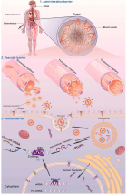
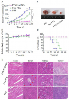
 Ultrasound imaging. (a) A schematic illustration of the basic principle of in vivo ultrasound imaging. First, the ultrasound contrast agent that encapsulates RNAi therapeutics is injected into animals, then high-frequency sound waves are sent and penetrated into the subjects, while the time intervals of subsequent reflection of sound waves is recorded by a transducer. The detected signals are converted and constructed into images. A coupling medium is usually used between the contact surface of transducer and subject for sound wave transmission. (b) Typical contrast agents for ultrasound, such as liposome, nanobubble, nanodroplet, and microbubble. (c) Ultrasound-triggered destruction of siRNA-incorporated microbubbles. Ultrasound could disrupt the contrast agents with high acoustic pressure to release RNAi agents when delivered to the blood vessels of the tumor region. Then the released RNAi agents enter into tumor cells to silence target gene." alt="jro-aid1035-g009"class="img-responsive img-rounded " style="cursor:pointer">
Figure 9:
Ultrasound imaging. (a) A schematic illustration of the basic principle of in vivo ultrasound imaging. First, the ultrasound contrast agent that encapsulates RNAi therapeutics is injected into animals, then high-frequency sound waves are sent and penetrated into the subjects, while the time intervals of subsequent reflection of sound waves is recorded by a transducer. The detected signals are converted and constructed into images. A coupling medium is usually used between the contact surface of transducer and subject for sound wave transmission. (b) Typical contrast agents for ultrasound, such as liposome, nanobubble, nanodroplet, and microbubble. (c) Ultrasound-triggered destruction of siRNA-incorporated microbubbles. Ultrasound could disrupt the contrast agents with high acoustic pressure to release RNAi agents when delivered to the blood vessels of the tumor region. Then the released RNAi agents enter into tumor cells to silence target gene.
Read Full Article HTML DOI: 10.29328/journal.jro.1001035 Cite this Article Read Full Article PDF
More Images
Similar Articles
-
Photon Absorption characteristics of some selected Enzyme Inhibitors used in Cancer Research in the Energy range 1 keV-100 GeVMustafa Recep Kaçal,Hüseyin Ali Karataş,Ferdi Akman*. Photon Absorption characteristics of some selected Enzyme Inhibitors used in Cancer Research in the Energy range 1 keV-100 GeV. . 2017 doi: 10.29328/journal.jro.1001009; 1: 060-068
-
Near Complete Response to 177Lu-PSMA-DKFZ-617 Therapy in a Patient with Metastatic Castration Resistant Prostate CancerMadhav Prasad Yadav,Sanjana Ballal a,Chandrasekhar Bal*. Near Complete Response to 177Lu-PSMA-DKFZ-617 Therapy in a Patient with Metastatic Castration Resistant Prostate Cancer. . 2017 doi: 10.29328/journal.jro.1001012; 1: 083-086
-
Hyperthermia and Breast cancer: A short reviewBora Uysal*. Hyperthermia and Breast cancer: A short review. . 2017 doi: 10.29328/journal.jro.1001011; 1: 079-082
-
Imaging modalities delivery of RNAi therapeutics in cancer therapy and clinical applicationsLoutfy H Madkour*. Imaging modalities delivery of RNAi therapeutics in cancer therapy and clinical applications. . 2021 doi: 10.29328/journal.jro.1001035; 5: 005-034
-
Anal cancer - impact of interstitial brachytherapyRegináčová K*,Kubecová M,Vránová J,Šejdová M. Anal cancer - impact of interstitial brachytherapy. . 2021 doi: 10.29328/journal.jro.1001034; 5: 001-004
-
Role of HRCT Thorax in preoperative assessment of RT-PCR COVID-19 negative oral cancer patientsRajesh A Kantharia*,Roopa Ganta,Shehnaz R Kantharia,Bhavin Bhupendra Shah,Rajinikant Mohan Vasava,Tushar Vishnuprasad Vaishnav. Role of HRCT Thorax in preoperative assessment of RT-PCR COVID-19 negative oral cancer patients. . 2020 doi: 10.29328/journal.jro.1001033; 4: 008-011.
-
8 Gy single dose radiotherapy for bone metastasis in COVID-19 pandemia period: ReviewBora Uysal*,Hakan Gamsız,Ferrat Dincoglan,Omer Sager,Selcuk Demiral ,Murat Beyzadeoglu. 8 Gy single dose radiotherapy for bone metastasis in COVID-19 pandemia period: Review. . 2020 doi: 10.29328/journal.jro.1001032; 4: 005-007.
-
Percentage of Positive Biopsy Cores Predicts Presence of a Dominant Lesion on MRI in Patients with Intermediate Risk Prostate CancerJason M Slater,William W Millard,Samuel M Randolph,Thomas J Kelly,David A Bush*. Percentage of Positive Biopsy Cores Predicts Presence of a Dominant Lesion on MRI in Patients with Intermediate Risk Prostate Cancer. . 2018 doi: 10.29328/journal.jro.1001025; 2: 073-079
-
Adaptive planning and toxicities of uniform scanning proton therapy for lung cancer patientsYuanshui Zheng*,Hardev Singh,Suresh Rana,Gary Larson,Prabhu Kiran,Lucius Doh,James Wong. Adaptive planning and toxicities of uniform scanning proton therapy for lung cancer patients. . 2018 doi: 10.29328/journal.jro.1001023; 2: 055-061
-
Incidental findings in traditional nuclear medicine practiceGiuseppe De Vincentis,Mariano Pontico,Maria Ricci,Viviana Frantellizzi*,Mauro Liberatore. Incidental findings in traditional nuclear medicine practice. . 2018 doi: 10.29328/journal.jro.1001019; 2: 036-040
Recently Viewed
-
Minimising Carbon Footprint in Anaesthesia PracticeNisha Gandhi and Abinav Sarvesh SPS*. Minimising Carbon Footprint in Anaesthesia Practice. Int J Clin Anesth Res. 2024: doi: 10.29328/journal.ijcar.1001025; 8: 005-007
-
On Friedman equation, quadratic laws and the geometry of our universeS Kalimuthu*. On Friedman equation, quadratic laws and the geometry of our universe. Int J Phys Res Appl. 2021: doi: 10.29328/journal.ijpra.1001041; 4: 048-050
-
Texture of Thin Films of Aluminum Nitride Produced by Magnetron SputteringStrunin Vladimir Ivanovich,Baranova Larisa Vasilievna*,Baisova Bibigul Tulegenovna. Texture of Thin Films of Aluminum Nitride Produced by Magnetron Sputtering. Int J Phys Res Appl. 2025: doi: 10.29328/journal.ijpra.1001106; 8: 013-016
-
KYAMOS Software - Mini Review on the Computer-Aided Engineering IndustryAntonis Papadakis*,Sofia Nikolaidou. KYAMOS Software - Mini Review on the Computer-Aided Engineering Industry. Int J Phys Res Appl. 2025: doi: 10.29328/journal.ijpra.1001105; 8: 010-012
-
The Theory of ElementsAbdellah El-Mourabet*. The Theory of Elements. Int J Phys Res Appl. 2025: doi: 10.29328/journal.ijpra.1001104; 8: 001-009
Most Viewed
-
Evaluation of Biostimulants Based on Recovered Protein Hydrolysates from Animal By-products as Plant Growth EnhancersH Pérez-Aguilar*, M Lacruz-Asaro, F Arán-Ais. Evaluation of Biostimulants Based on Recovered Protein Hydrolysates from Animal By-products as Plant Growth Enhancers. J Plant Sci Phytopathol. 2023 doi: 10.29328/journal.jpsp.1001104; 7: 042-047
-
Sinonasal Myxoma Extending into the Orbit in a 4-Year Old: A Case PresentationJulian A Purrinos*, Ramzi Younis. Sinonasal Myxoma Extending into the Orbit in a 4-Year Old: A Case Presentation. Arch Case Rep. 2024 doi: 10.29328/journal.acr.1001099; 8: 075-077
-
Feasibility study of magnetic sensing for detecting single-neuron action potentialsDenis Tonini,Kai Wu,Renata Saha,Jian-Ping Wang*. Feasibility study of magnetic sensing for detecting single-neuron action potentials. Ann Biomed Sci Eng. 2022 doi: 10.29328/journal.abse.1001018; 6: 019-029
-
Pediatric Dysgerminoma: Unveiling a Rare Ovarian TumorFaten Limaiem*, Khalil Saffar, Ahmed Halouani. Pediatric Dysgerminoma: Unveiling a Rare Ovarian Tumor. Arch Case Rep. 2024 doi: 10.29328/journal.acr.1001087; 8: 010-013
-
Physical activity can change the physiological and psychological circumstances during COVID-19 pandemic: A narrative reviewKhashayar Maroufi*. Physical activity can change the physiological and psychological circumstances during COVID-19 pandemic: A narrative review. J Sports Med Ther. 2021 doi: 10.29328/journal.jsmt.1001051; 6: 001-007

HSPI: We're glad you're here. Please click "create a new Query" if you are a new visitor to our website and need further information from us.
If you are already a member of our network and need to keep track of any developments regarding a question you have already submitted, click "take me to my Query."


