Abstract
Research Article
Inter-Observer Variability of a Commercial Patient Positioning and Verification System in Proton Therapy
Yuanshui Zheng* and Xiaoning Ding
Published: 06 February, 2017 | Volume 1 - Issue 1 | Pages: 031-038
Purpose:Accurate patient positioning is crucial in radiation therapy. To fully benefit from the preciseness of proton therapy, image guided patient positioning and verification system is typically utilized in proton therapy. The purpose of this study is to evaluate the inter-observer variability of image alignment using a commercially available patient positioning and verification system in proton therapy.
Methods:The VeriSuite patient positioning and verification system (MedCom GmbH, Darmstadt, Germany) provides a six degrees of freedom correction vector by registering two orthogonal x-ray images to digitally reconstructed radiograph (DRR) images that are rendered in real time from the planning computed tomography (CT) images. Six cases of various disease sites, including brain, head & neck, lung, prostate, pelvis, and bladder, were used in this study. For each case, the planning CT images and a daily orthogonal x-ray portal image pair were loaded into the VeriSuite system. The same set of x-ray images and CT images for each case were reviewed and aligned separately by each of the 10 radiation therapist, following the clinical procedure for the corresponding disease site. The resulting correction vectors were then recorded and analyzed.
Results:Our study shows that the inter-observer variation (One standard deviation) in image alignment using the VeriSuite system ranged from 1.2 to 2.0 mm for translational correction and from 0.6 to 1.3 degrees for rotational correction for the six cases. The use of fiducial markers for prostate patient alignment achieved the least inter-observer variation while the bladder case produced the largest.
Conclusions:Inter-observer variation in image alignment could be relatively large, depending on the complexity of patient anatomy, image alignment approach, and user experience and software limitations. Automatic registration and fiducial markers could potentially be used to align patient more accurately and consistently. To ensure adequate tumor coverage in proton therapy, inter-observer variability in patient alignment should be carefully evaluated and accounted for in patient setup uncertainty analysis and treatment planning margin determination.
Read Full Article HTML DOI: 10.29328/journal.jro.1001004 Cite this Article Read Full Article PDF
Keywords:
Inter-observer variability; Image alignment; Proton therapy
References
- Engelsman M, Schwarz M, Dong L. Physics controversies in proton therapy. Semin Radiat Oncol. 2013; 23: 88-96. Ref.: https://goo.gl/4gbQgV
- Rosen L. Everything is changing in oncology and proton therapy. J Proton Ther. 2015; 1: 111. Ref.: https://goo.gl/zwd392
- Yin F, Wong J, Balter J, Benedict S, Craig J, et al. The role of in-room kV X-ray imaging for patient setup and target localization. Report of AAPM Task Group. 2009; 104. Ref.: https://goo.gl/rYpMVS
- De Los Santos J, Popple R, Agazaryan N, Bayouth JE, Bissonnette JP, et al. Image guided radiation therapy (IGRT) technologies for radiation therapy localization and delivery. Int J Radiat Oncol Biol Phys. 2013; 87: 33-45.Ref.: https://goo.gl/7QP8CU
- Fuss M, Salter BJ, Cavanaugh SX, Fuss C, Sadeghi A, et al. Daily ultrasound-based image-guided targeting for radiotherapy of upper abdominal malignancies. Int J Radiat Oncol Biol Phys. 2004; 59: 1245-1256. Ref.: https://goo.gl/iHRfcE
- Molloy JA, Chan G, Markovic A, McNeeley S, Pfeiffer D, et al. Quality assurance of U.S.-guided external beam radiotherapy for prostate cancer: report of AAPM Task Group 154. Med Phys. 2011; 38: 857-871. Ref.: https://goo.gl/1gjnSQ
- Wooten HO, Rodriguez V, Green O, Kashani R, Santanam L, et al. Benchmark IMRT evaluation of a Co-60 MRI-guided radiation therapy system. Radiother Oncol. 2015; 114: 402-405. Ref.: https://goo.gl/eFrX0L
- Oelfke U, Tucking T, Nill S, Seeber A, Hesse B, et al. Linac-integrated kV-cone beam CT: technical features and first applications. Med Dosim. 2006; 3: 62-70. Ref.: https://goo.gl/XHfpFn
- Morin O, Gillis A, Chen J, Aubin M, Bucci MK 3rd, et al. Megavoltage cone-beam CT: system description and clinical applications. Med Dosim. 2006; 31: 51-61. Ref.: https://goo.gl/WqIuHI
- Bert C, Metheany KG, Doppke KP, Taghian AG, Powell SN, et al. Clinical experience with a 3D surface patient setup system for alignment of partial-breast irradiation patients. Int J Radiat Oncol Biol Phys. 2006; 64: 1265-1274. Ref.: https://goo.gl/RAah6i
- Fattori G, Riboldi M, Pella A, Peroni M, Cerveri P, et al. Image guided particle therapy in CNAO room 2: implementation and clinical validation. Phys Med. 2015; 31: 9-15. Ref.: https://goo.gl/jxDrtO
- Depauw N, Batin E, Daartz J, Rosenfeld A, Adams J, et al. A novel approach to postmastectomy radiation therapy using scanned proton beams. Int J Radiat Oncol Biol Phys. 2015; 91: 427-434. Ref.: https://goo.gl/ffCgk0
- Enke C, Ayyangar KM, Saw CB, Zhen W, Thompson RB, et al. Inter-observer variation in prostate localization utilizing BAT. Int J Radiat Oncol Biol Phys. 2002; 54: 269. Ref.: https://goo.gl/ahKfKs
- Aubin M, Liu Y, Langen K, Shinohara K, Anezinos C, et al. Set-up verification using portal images of implanted prostate markers: an inter-observer study. Int J Radiat Oncol Biol Phys. 2002; 54: 269-270. Ref.: https://goo.gl/xSUqNI
- Ullman KL, Ning H, Susil RC, Ayele A, Jocelyn L, et al. Intra- and inter-radiation therapist reproducibility of daily isocenter verification using prostatic fiducial markers. Radiat Oncol. 2006; 1: 2. Ref.: https://goo.gl/FLxAde
- Court LE, Dong L, Taylor N, Ballo M, Kitamura K, et al. Evaluation of a contour-alignment technique for CT-guided prostate radiotherapy: an intra- and interobserver study. Int J Radiat Oncol Biol Phys. 2004; 59: 412-418. Ref.: https://goo.gl/VSUDZv
- Court LE, Allen A, Tishler R. Evaluation of the precision of portal-image-guided head-and-neck localization: an intra- and interobserver study. Med Phys. 2007; 34: 2704-2707. Ref.: https://goo.gl/tIx8VX
- Guckenberger M, Sweeney RA, Wilbert J, Krieger T, Richter A, et al. Image-guided radiotherapy for liver cancer using respiratory-correlated computed tomography and cone-beam computed tomography. Int J Radiat Oncol Biol Phys. 2008; 71: 297-304. Ref.: https://goo.gl/3buPTa
- Commission IE. IEC 61217 radiotherapy equipment-co-ordinates, movements and scales. International Electrotechnical Commission. 2011. Ref.: https://goo.gl/q0LxHM
- Pella A, Riboldi M, Tagaste B, Bianculli D, Desplanques M, et al. Commissioning and quality assurance of an integrated system for patient positioning and setup verification in particle therapy. Technol Cancer Res T. 2014; 13: 303-314. Ref.: https://goo.gl/Ece8Ao
- Walter S. Automatic patient alignment in six degrees of freedom for particle beam treatment. PTCOG; 2007 May 18-23; Wanjie, Shandong, China.
- Sejpal SV, Amos RA, Bluett JB, Levy LB, Kudchadker RJ, et al. Dosimetric changes resulting from patient rotational setup errors in proton therapy prostate plans. Int J Radiat Oncol Biol Phys. 2009; 75: 40-48. Ref.: https://goo.gl/w6Z32h
- Rana S, Zhang Y, Larson G, Vargas C, Dunn M, et al. Investigating dosimetric effect of rotational setup errors in IMPT planning of synchronous bilateral lung cancer. Int J Cancer Ther Oncol. 2015; 3. Ref.: https://goo.gl/99xLw5
- Fuss M, Cavanaugh SX, Fuss C, Cheek DA, Salter BJ. Daily stereotactic ultrasound prostate targeting: inter-user variability. Technol Cancer Res Treat. 2003; 2: 161-170. Ref.: https://goo.gl/QmxFHr
Figures:
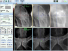
Figure 1

Figure 2
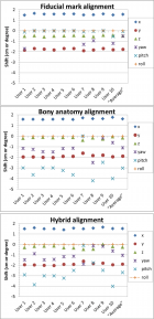
Figure 3
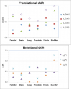
Figure 4
Similar Articles
-
Inter-Observer Variability of a Commercial Patient Positioning and Verification System in Proton TherapyYuanshui Zheng*,Xiaoning Ding. Inter-Observer Variability of a Commercial Patient Positioning and Verification System in Proton Therapy. . 2017 doi: 10.29328/journal.jro.1001004; 1: 031-038
-
Biological dose calculation using variable RBE in Single- and Multi-field Optimization IMPT plans for 3 Brain Tumor patientsRyosuke Kohno*,Wenhua Cao,Pablo Yepes,Xuemin Bai,Falk Poenisch,David R Grosshans,Tetsuo Akimoto,Radhe Mohan. Biological dose calculation using variable RBE in Single- and Multi-field Optimization IMPT plans for 3 Brain Tumor patients. . 2019 doi: 10.29328/journal.jro.1001027; 3: 001-007
-
Adaptive planning and toxicities of uniform scanning proton therapy for lung cancer patientsYuanshui Zheng*,Hardev Singh,Suresh Rana,Gary Larson,Prabhu Kiran,Lucius Doh,James Wong. Adaptive planning and toxicities of uniform scanning proton therapy for lung cancer patients. . 2018 doi: 10.29328/journal.jro.1001023; 2: 055-061
Recently Viewed
-
Metastatic Brain Melanoma: A Rare Case with Review of LiteratureNeha Singh,Gaurav Raj,Akshay Kumar,Deepak Kumar Singh,Shivansh Dixit,Kaustubh Gupta*. Metastatic Brain Melanoma: A Rare Case with Review of Literature. J Radiol Oncol. 2025: doi: ; 9: 050-053
-
Depression as a civilization-deformed adaptation and defence mechanismBohdan Wasilewski*,Olha Yourtsenyuk,Eugene Egan. Depression as a civilization-deformed adaptation and defence mechanism. Insights Depress Anxiety. 2020: doi: 10.29328/journal.ida.1001013; 4: 008-011
-
Drinking-water Quality Assessment in Selective Schools from the Mount LebanonWalaa Diab, Mona Farhat, Marwa Rammal, Chaden Moussa Haidar*, Ali Yaacoub, Alaa Hamzeh. Drinking-water Quality Assessment in Selective Schools from the Mount Lebanon. Ann Civil Environ Eng. 2024: doi: 10.29328/journal.acee.1001061; 8: 018-024
-
Rapid Microbial Growth in Reusable Drinking Water BottlesQishan Liu*,Hongjun Liu. Rapid Microbial Growth in Reusable Drinking Water Bottles. Ann Civil Environ Eng. 2017: doi: 10.29328/journal.acee.1001007; 1: 055-062
-
Beneficial effects of a ketogenic diet in a woman with Charcot-Marie-Tooth diseaseElvira Rostanzo,Anna Maria Aloisi*. Beneficial effects of a ketogenic diet in a woman with Charcot-Marie-Tooth disease. Arch Food Nutr Sci. 2022: doi: 10.29328/journal.afns.1001040; 6: 068-072
Most Viewed
-
Evaluation of Biostimulants Based on Recovered Protein Hydrolysates from Animal By-products as Plant Growth EnhancersH Pérez-Aguilar*, M Lacruz-Asaro, F Arán-Ais. Evaluation of Biostimulants Based on Recovered Protein Hydrolysates from Animal By-products as Plant Growth Enhancers. J Plant Sci Phytopathol. 2023 doi: 10.29328/journal.jpsp.1001104; 7: 042-047
-
Sinonasal Myxoma Extending into the Orbit in a 4-Year Old: A Case PresentationJulian A Purrinos*, Ramzi Younis. Sinonasal Myxoma Extending into the Orbit in a 4-Year Old: A Case Presentation. Arch Case Rep. 2024 doi: 10.29328/journal.acr.1001099; 8: 075-077
-
Feasibility study of magnetic sensing for detecting single-neuron action potentialsDenis Tonini,Kai Wu,Renata Saha,Jian-Ping Wang*. Feasibility study of magnetic sensing for detecting single-neuron action potentials. Ann Biomed Sci Eng. 2022 doi: 10.29328/journal.abse.1001018; 6: 019-029
-
Pediatric Dysgerminoma: Unveiling a Rare Ovarian TumorFaten Limaiem*, Khalil Saffar, Ahmed Halouani. Pediatric Dysgerminoma: Unveiling a Rare Ovarian Tumor. Arch Case Rep. 2024 doi: 10.29328/journal.acr.1001087; 8: 010-013
-
Physical activity can change the physiological and psychological circumstances during COVID-19 pandemic: A narrative reviewKhashayar Maroufi*. Physical activity can change the physiological and psychological circumstances during COVID-19 pandemic: A narrative review. J Sports Med Ther. 2021 doi: 10.29328/journal.jsmt.1001051; 6: 001-007

HSPI: We're glad you're here. Please click "create a new Query" if you are a new visitor to our website and need further information from us.
If you are already a member of our network and need to keep track of any developments regarding a question you have already submitted, click "take me to my Query."


